
A Clearer Path Through Operative Hysteroscopy: What Patients and Care Teams Should Know in 2026
Operative hysteroscopy is a procedure that allows a clinician to look inside the uterus and, when appropriate, treat certain findings during the same visit. It may be discussed when symptoms, imaging, or office evaluation suggest a concern within the uterine cavity. For example, learning more about fibroids symptoms may help patients understand why further evaluation is sometimes recommended. Every patient’s situation is different. The reason for hysteroscopy, the setting where it is performed, the type of anesthesia or pain control used, and the recovery plan should be based on an individual clinical assessment. Patients should rely on guidance from their own clinician for instructions before and after a procedure.
What Is Operative Hysteroscopy?
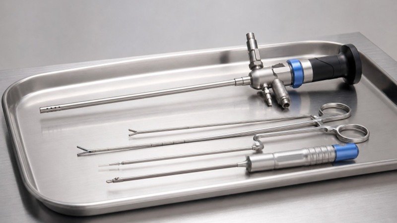
Hysteroscopy involves placing a narrow instrument with a camera through the vagina and cervix to view the inside of the uterus. A diagnostic hysteroscopy is primarily used to examine the uterine cavity. An operative hysteroscopy includes the use of specialized instruments to remove, cut, sample, or otherwise treat selected tissue when clinically appropriate. Because the clinician can directly view the uterine cavity, hysteroscopy may allow evaluation and treatment without an abdominal incision. The appropriate approach depends on the suspected condition, the patient’s medical history, and the characteristics of the tissue being evaluated.
When It May Be Used
A clinician may consider operative hysteroscopy after symptoms, ultrasound findings, saline infusion sonography, or another evaluation indicate a possible issue inside the uterus. Common reasons for further discussion can include:
- Uterine polyps.
- Selected submucosal fibroids, which extend into the uterine cavity.
- Scar tissue or intrauterine adhesions.
- Retained tissue after pregnancy, when clinically indicated.
- Selected causes of abnormal uterine bleeding.
Not every polyp, fibroid, or bleeding concern can be managed hysteroscopically. Size, location, number of findings, anatomy, pathology results, and other medical considerations can affect whether this approach is suitable.
How the Procedure Works
While the details differ by patient and facility, operative hysteroscopy commonly follows a structured process:
- The care team confirms the treatment plan, health history, consent, and relevant test results.
- The patient receives the planned pain control, sedation, or anesthesia.
- The clinician gently places the hysteroscope through the cervix.
- Sterile fluid is used to expand the uterine cavity and support visualization.
- The clinician examines the cavity and treats the identified target when appropriate.
- The team documents key details and monitors the patient in recovery.
In some cases, the ability to see and treat a finding in the same procedure can reduce the need for separate diagnostic and treatment visits. However, a clinician may decide to stop, reschedule, obtain additional information, or recommend another approach if the planned treatment cannot be completed safely.
Why Clear Visualization Matters
A clear view of the uterine cavity helps the clinician identify the target tissue, distinguish it from surrounding structures, and guide instrument movement. Bleeding, bubbles, tissue fragments, and cloudy fluid can make visualization more difficult.
Pausing Can Be Part of Safe Care
If a small polyp or other finding is not clearly visible, the clinician may pause to improve the view and reassess the cavity. This deliberate approach supports informed procedural decisions rather than proceeding without a reliable visual field.
Fluid Management and Safety
Fluid used during hysteroscopy expands the uterine cavity, creating space for visualization. The care team tracks the amount of fluid entering and leaving the procedure field because excessive fluid absorption can lead to serious complications. Important fluid-management practices may include recording the type of fluid, tracking inflow and outflow, monitoring the patient’s condition, recognizing changes in visualization or pressure, and documenting the final fluid balance. Facilities should follow their established protocols and clinical policies.
Preparing for the Procedure
Preparation instructions vary according to the planned setting and anesthesia plan. Before the procedure, patients may be asked to:
- Share a complete list of medications, vitamins, and supplements.
- Discuss allergies, pregnancy concerns, prior surgeries, and anesthesia history.
- Follow instructions about eating, drinking, and medication use.
- Arrange transportation if sedation or anesthesia is planned.
- Ask when work, exercise, bathing, and sexual activity may resume.
General online information cannot replace instructions from the treating clinician. If a patient is uncertain about preparation steps, contacting the care team before the appointment is appropriate.
Questions to Ask Before Treatment
Shared decision-making begins with understanding the goal of the procedure. Useful questions may include:
- What finding is this procedure intended to evaluate or treat?
- Are there alternatives to hysteroscopy in this situation?
- What type of pain control or anesthesia is planned?
- How will fluid balance be monitored?
- What happens if all planned treatment cannot be completed?
- Will the removed tissue be sent for laboratory testing?
- Which symptoms should prompt an urgent call after the procedure?
Recovery and Follow-Up
Some patients experience mild cramping, light bleeding or spotting, discharge, or temporary fatigue after sedation or anesthesia. Recovery experiences vary based on the procedure performed and the individual patient.
Patients should receive written discharge instructions that explain medication use, activity guidance, follow-up plans, and when to seek medical advice. Heavy bleeding, severe or worsening pain, fever, fainting, breathing difficulty, or other concerning symptoms should be reported promptly to the treating team or addressed through urgent medical care.
What Care Teams Should Review
Consistent communication and preparation support an organized procedure experience. Key items for care teams to review include:
- Patient identity, consent, planned procedure, and treatment site.
- Equipment readiness, sterilization status, and availability of backup instruments.
- Anesthesia planning, emergency supplies, and escalation procedures.
- Fluid monitoring responsibilities and documentation.
- Specimen handling, labeling, and laboratory processes.
- Recovery instructions and follow-up arrangements.
Conclusion
Operative hysteroscopy can serve both diagnostic and treatment purposes for selected conditions within the uterine cavity. Thoughtful patient selection, clear visualization, careful fluid management, coordinated teamwork, and individualized follow-up are all important parts of the process. Patients who understand the treatment plan are often better prepared to participate in decisions about their care and know what to expect before, during, and after the procedure. Open communication with the healthcare team, realistic expectations about recovery, and adherence to post-procedure instructions can also support a smoother healing process. While every patient’s situation is different, careful planning and personalized care help reduce uncertainty and improve the overall treatment experience. With appropriate evaluation and follow-up, operative hysteroscopy can be an effective option for addressing a range of uterine conditions while supporting both short-term recovery and long-term reproductive or gynecologic health.


